Designing dual binding site acetylcholinesterase inhibitors ……
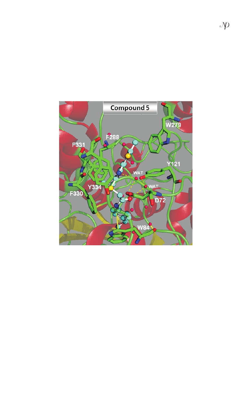
formed π-π stacking interaction with the W84 of AChE enzyme. In addition, two
water mediated hydrogen bonding interactions were also observed between (i)
the carbonyl oxygen (C=O) of compound 5 and hydroxyl group of Y121 through
water (WAT1157), and (ii) second water (WAT1350) mediated hydrogen bonding
interaction was observed between one of the nitrogen atom of thienopyrimidine
ring of compound 5 and carboxylate group of D72, in the middle of the gorge site
(CS) of the AChE enzyme as shown in Figure 5.
fig. 5: molecular docking derived binding pose of the lead compound 5 in the dual
binding site (cS and PaS) of achE enzyme. the inhibitor is shown as ball and stick
model in the surface representation of the enzyme. Water compounds are shown as
red dotted spheres. Hydrogen bonding is shown as dotted lines (yellow color) and
π-π interaction by straight line (yellow color). GOLD software was used to derive
the binding mode and the picture was generated from PymoL software.
conclusion
In summary, we report here a novel FBDD approach to generate new dual binding
siteAChEIs followed byADMET and docking analysis.The present study revealed
that the dissection of co-crystallized inhibitors is an interesting approach to select
the best fragments in terms of binding efficiencies and synthetic feasibility. Rules
similar to those used in RECAP were applied in fragment generation process
and suitable fragments that bound simultaneously to CS and PAS of the AChE
binding site were obtained to satisfy the primary requirement of this strategy.
Int. J. Bioinformatics and Biological Sci.: v.2 n.1&2, p. 111-126. March & June 2014
123