Gross and Biometrical studies of Placentome in Goat
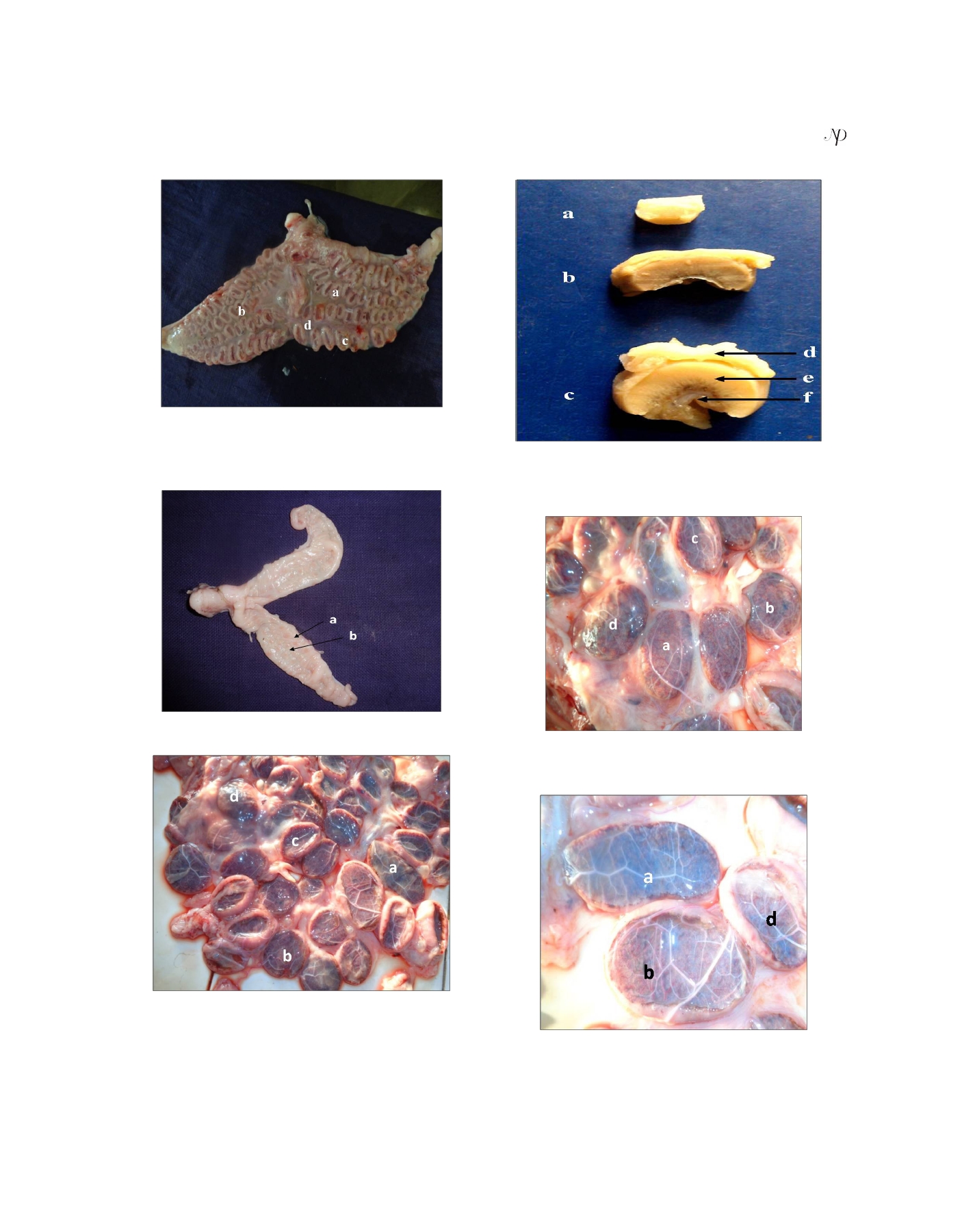
Figure 1: Photograph of goat uterus at 90 days of pregnancy
showing (a) gravid horn, (b) non gravid horn, (c) placentome
(concave shape with thick margin) and (d) inter placentomal
Figure 4. Photograph showing different stage of placentome in
area.
relation of attachment either uterine wall or chorionic epithelium
(a) early pregnancy (b) mid pregnancy (c) late pregnancy (d)
uterine wall (e) caruncle (f) chorionic membrane.
Figure 2: Photograph of goat uterus at 23 days of pregnancy
showing (a) caruncle (b) intercaruncular area.
Figure 5: photograph of goat uterus at 134 days of pregnancy
showing (a) elliptical shape (b) flat shape (c) concave
plancentome with thin margin (d) convex plancentome
Figure 3: Photograph of goat uterus at 134 days of pregnancy
showing (a) elliptical shape placentome, (b) flat placentome (c)
placentome (concave shape with thin margin) and (d) placentome
(convex shape).
Figure 6: photograph of goat uterus at 134 days of pregnancy
showing (a) elliptical shape (b) flat shape (d) convex plancentome
Journal of Animal Research: v.5 n.2. June 2015
253